
|
Fe55 Xray Source |
|
For this Xray source the responsible person is Simon Tulloch. No
one is to use it without his supervision. The source is very low activity
(below the licensing threshold). When kept in its storage cryostat the Xrays
emitted by the source are fully blocked by the cryostat walls.
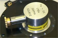
This 1 MBequerel Fe55 source was purchased in Jan 2004. Product code
IERB11784. Supplied by Nucliber in Madrid (Fax 091 539 4330).
It is the highest activity source that we can hold without having to pay
licensing fees. The geometry of the source gives approximately 1000 xray
events per second per square centimetre in a thinned CCD. Exposures of approximately
20s are required in order to obtain enough events for measurement of CTE
and gain. The half life of the Fe55 isotope is approximately 2.6 years.
The source is deposited on a small coin like copper disc and overcoated
with nickel. This disc is mounted on a vacuum rotary feedthrough where it
can be turned to face the detector to make the exposure. When turned away
the detector receives no Xrays. In this system the source is operated in
a vacuum. This avoids the need for a Beryllium window (transparent to soft
Xrays) which is an extremely hazardous material. For safety purposes we
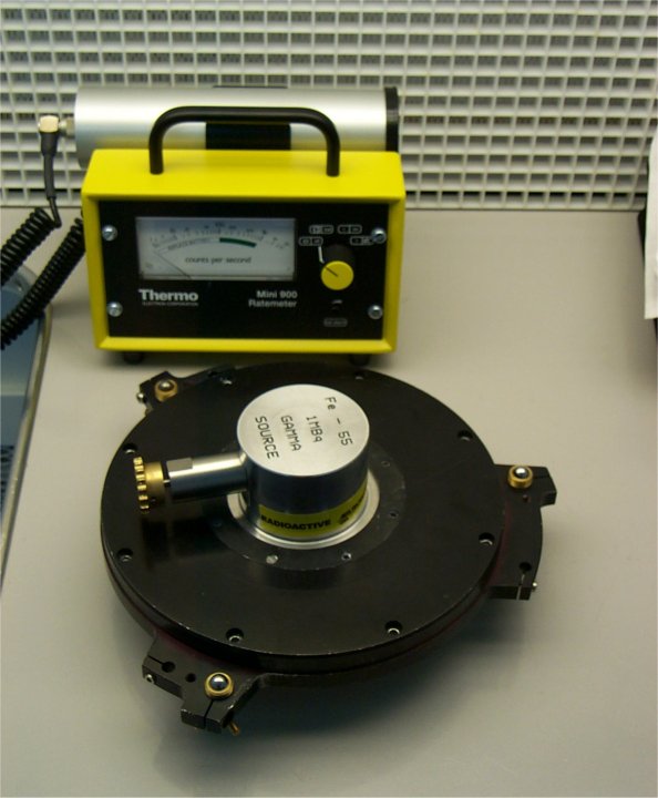
also have a soft Xray scintillation monitor : a Mini Instruments 900-44B.
This is supplied by ThermoElectron corporation in Reading UK (fax 0044 1189
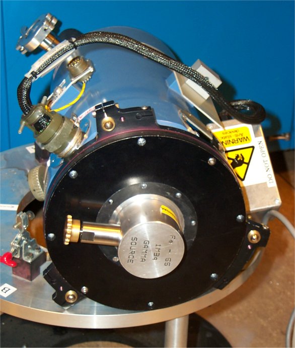
712 835). When not used the source is stored under vacuum inside a detector
cryostat.
The source emits X rays at three energies. The emission is caused by the
inner electron of the Fe55 isotope being captured by the nucleus, transforming
it into Manganese. By far the most intense of these emissions is at 5.899KeV
(the so called Mn KAlpha line), but there are weaker peaks at 6.490KeV (Mn
KBeta) and 4.12KeV (KAlpha escape). When these Xrays are absorbed by silicon
they produce large photoelectron events:
Mn KAlpha gives 1620 electrons, Mn KBeta 1778 electrons and the KAlpha
escape 1133electrons. Occaisonally, the absorbed Xray photon is re-emitted
(fluoresced) by the silicon of the detector and is reabsorbed later where
it produces a photo-electron event of 487e. All of these X rays are easily
attenuated: 1mm of aluminium reduces the 6KeV flux by a factor of 1 million.
The cryostat walls are 3mm thick.
|
|
| Source and Monitor |
Source mounted
on CCD Camera |
Gain (i.e. electrons per ADU) measurements are very staightforward and
accurate using this system. The Xray images are histogrammed and the bias
and Mn KAlpha pixels are identified.The difference between these peaks
corresponds to 1620e. Horizontal charge transfer efficiency (HCTE)can be
calculated by comparing histograms taken from pixels at the left and right
hand sides of the image. A poor CTE chip will show lower Xray events at
the side of the image furthest from the readout amplifier. Likewise, comparing
histograms of pixels at top and bottom of the image gives the Vertical charge
transfer efficiency (VCTE).
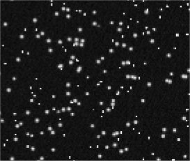
Most Xrays pass right through a thinned CCD. The 20% or so that are stopped
are absorbed all depths throughout the silicon. Most of the charge that
is generated is shared between several pixels ; so called split events.
A very small percentage , however, create 'single pixel events' and it is
these that are useful diagnostically. The image below shows just how scarce
these single pixel events are.
|
| A small section of an Xray image. |
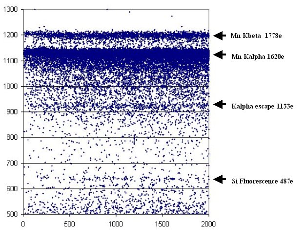
The plot below is derived from an Xray image taken with the EEV10 camera
( which contains a CCD4280 device). The pixel data was fed through a filter
program to extract only the single pixel X ray events. The program gave
an output consisting of the X and Y coordinates as well as the height in
ADU of each of these events. This plot shows the event height along the
y axis and the column number of the event along the x axis. The bias level
was 435ADU.
Simon Tulloch
April 2004